Why Wisdom Teeth X-Rays Matter Before Extraction Planning
Wisdom teeth can be difficult to understand from symptoms alone. You might feel pressure at the back of the jaw, notice swelling around the gum, or have no discomfort at all while a tooth is still developing under the surface.
That is why getting a wisdom teeth x-ray is often an important first step before extraction planning – Adelaide patients can discuss their options with our experienced dental team at Kensington Dental Care. Imaging helps your dentist see what cannot be confirmed by looking inside the mouth, including the tooth’s position, root shape, nearby teeth, jawbone, and important structures such as nerves and sinuses.
At Kensington Dental Care, we assess wisdom tooth growth carefully before recommending the next step. Some wisdom teeth can be monitored. Others may need removal because they are impacted, infected, affecting nearby teeth, or difficult to clean properly. The right plan depends on your individual situation, not just whether a wisdom tooth is present.
Why an X-Ray Matters Before Wisdom Tooth Removal
A wisdom tooth sits at the very back of the mouth, where visibility is limited. Even when part of the tooth has come through the gum, the most important information may still be hidden under the surface.
Dental imaging can help your dentist assess:
- Whether the wisdom tooth has enough room to come through
- The angle of the tooth and whether it is impacted
- The shape and direction of the roots
- Whether decay or gum issues are affecting the wisdom tooth or the tooth in front
- How close the tooth appears to nerves, sinuses, or other anatomy
- Whether a straightforward extraction or more detailed surgical planning may be needed
This information supports a more informed conversation about your options. If you are planning wisdom teeth removal, an x-ray can help your dentist explain what they can see, what risks need to be considered, and whether further imaging would help.
What Dentists Look For on a Wisdom Teeth X-Ray
A wisdom teeth assessment at our Adelaide practice usually combines a clinical examination with imaging. Your dentist will check your gums, bite, jaw movement, symptoms, medical history, and oral hygiene around the back molars.
The x-ray then adds context. It can show whether the wisdom tooth is upright, tilted forward, angled backwards, lying horizontally, or trapped below the gum. This matters because a wisdom tooth that is pushing into the second molar may be more likely to cause food trapping, decay, gum inflammation, or damage to the neighbouring tooth.
Your dentist may also look at whether the tooth is fully covered by bone, partly through the gum, or close to the roots of nearby teeth. In some cases, the x-ray may show early signs of cyst formation around an unerupted tooth. Not every finding means the tooth must be removed immediately, but it does help guide a careful plan.
OPG X-Rays: The Panoramic First Look
An OPG, also called a panoramic x-ray, is commonly used as a first look at wisdom teeth. It captures a broad view of the mouth and jaws in one image, making it useful when your dentist needs to understand all four wisdom teeth and their relationship to the surrounding teeth.
For many people, an OPG provides enough information to decide whether wisdom teeth should be monitored, removed in the dental chair, or assessed in more detail. It can show the position of unerupted teeth, the general direction of the roots, and whether a wisdom tooth is sitting close to the tooth in front.
An OPG is a two-dimensional image, so it has limits. It can suggest that a lower wisdom tooth is near the nerve canal, but it may not show the exact three-dimensional relationship between the roots and nerve. If that relationship could change the treatment plan, your dentist may recommend additional imaging.
When CBCT Imaging May Be Recommended
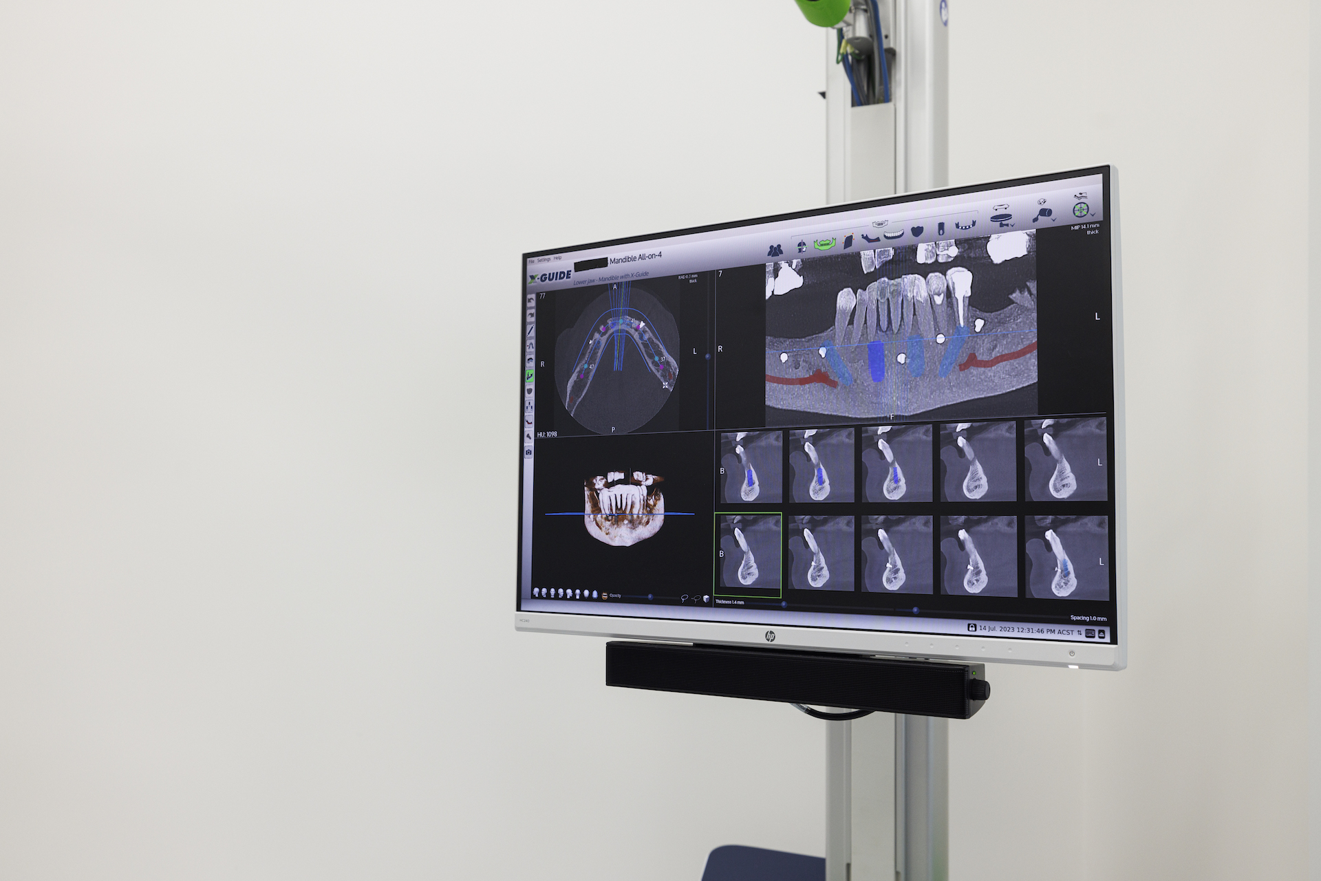
CBCT stands for cone beam computerised tomography. Unlike an OPG, a CBCT scan creates a three-dimensional image of the teeth, bone, and surrounding anatomy.
CBCT is not needed for every wisdom tooth. It may be considered when the OPG suggests a more complex situation, such as:
- Lower wisdom tooth roots that appear close to the inferior dental nerve
- Unusual root shape or unclear root position
- Deep impaction within the jawbone
- A tooth that sits close to the sinus
- Complex surgical extraction planning
- Cases where three-dimensional information may change how the extraction is planned
The aim is not to order more imaging than necessary. It is to gather the right information for the level of risk and complexity. When planning impacted wisdom teeth treatment, the value of imaging is in matching the plan to the anatomy.
Impacted Wisdom Teeth and Why Position Changes the Plan
An impacted wisdom tooth is one that cannot fully come through into the mouth. It may be blocked by bone, gum tissue, or the tooth in front. Some impacted wisdom teeth cause pain or swelling, while others are found during routine dental imaging.
Impacted teeth can create problems when they are hard to clean or when they place pressure on nearby structures. They may contribute to:
- Gum infection around a partly erupted tooth
- Food trapping and bad taste near the back molars
- Tooth decay on the wisdom tooth or second molar
- Pain, swelling, or jaw stiffness
- Cysts around an unerupted tooth
- Damage to nearby teeth
The angle and depth of the tooth matter. A shallow, partly erupted wisdom tooth may be managed differently from a deeply impacted lower tooth with roots close to the nerve canal. Imaging helps your dentist explain why one case may be relatively simple while another needs more detailed surgical planning.
If discomfort is your main concern, our guide to wisdom teeth pain relief may help you understand short-term care while you arrange a proper assessment.
Nerve Proximity: Why Lower Wisdom Teeth Need Careful Assessment
Lower wisdom teeth sit near the inferior dental nerve, which runs through the lower jaw. This nerve supplies feeling to parts of the lower lip, chin, and teeth.
Most wisdom tooth extractions do not involve unusual nerve concerns, but some lower teeth sit very close to the nerve canal. On a two-dimensional x-ray, the roots and nerve canal may appear to overlap. In those cases, three-dimensional imaging may help clarify whether the roots are beside, above, below, or wrapped around the canal.
This does not mean every close-looking tooth is dangerous or that every patient needs advanced imaging. It means your dentist should assess the x-ray in context and explain whether the finding changes the plan. Sometimes the next step is a CBCT scan. Sometimes it is a closer look at the images with your dentist. Sometimes the OPG and clinical examination provide enough information.
Why Symptoms Alone Can Be Misleading
When planning relies too heavily on symptoms, the picture can be misleading.
A tooth can be painful because it is partly erupted and the surrounding gum is inflamed. Another tooth may cause no obvious discomfort but still be positioned in a way that damages the second molar. A third may look visible in the mouth but have curved roots that affect how it should be removed.
X-rays help reduce uncertainty by showing the factors that shape the plan. They also help your dentist talk through reasonable options, including monitoring, cleaning support, removal under local anaesthetic in the dental chair, or more complex planning for surgical removal.
What your x-rays show also informs the anaesthetic conversation, which is not an all-or-nothing choice. Straightforward cases can often be treated in the chair under local anaesthetic. If you would rather not be awake but a hospital admission is not needed, IV sedation can be provided in the chair with a specialist anaesthetist present throughout. General anaesthetic is always undertaken in an SA Health licensed and accredited hospital or day surgery.
Dr Evan Jones is an experienced general dentist who consults and treats at a number of accredited hospitals — so if a general anaesthetic is the right pathway for you, the dentist who assessed and planned your case is the one who carries it out. If general anaesthetic suits your case, our guide to wisdom teeth removal under general anaesthetic cost explains the factors that shape the fee. Your dentist will discuss what is appropriate for your case after assessment.
Do You Need to See an Oral and Maxillofacial Surgeon?
Most wisdom tooth cases, particularly for patients who are fit and healthy, do not require referral to an oral and maxillofacial surgeon. Complex cases, or cases that would benefit from specialist management, are referred appropriately. Safety and the best patient outcome determine whether Dr Evan Jones performs the surgery or recommends specialist referral.
What Happens During a Wisdom Teeth Removal Consultation
When Adelaide patients book a wisdom teeth removal consultation with our team, the appointment is designed to give you a clear understanding of what is happening and what your options are.
Your appointment may include:
- A discussion about your symptoms and concerns
- A review of your dental and medical history
- An examination of the wisdom tooth area
- Dental imaging where clinically appropriate
- An explanation of whether your wisdom teeth can be monitored or need treatment
- A treatment plan with the expected approach, timing, and aftercare
We encourage you to ask questions. Many patients want to know whether all four wisdom teeth need attention, whether one tooth can be treated first, how recovery usually works, and whether the x-ray shows any added complexity. These are all reasonable points to raise during your consultation.
If you are still learning about these teeth, our article on adult wisdom teeth explains when they commonly appear and why not everyone has the same number.
Do Wisdom Teeth Always Need to Be Removed?
No. The presence of wisdom teeth alone does not automatically mean they need to be extracted.
Some wisdom teeth come through fully, sit in a useful position, and can be cleaned properly. Others stay under the gum without causing immediate problems and may be monitored with regular check-ups. Your dentist will consider your age, symptoms, oral hygiene, x-ray findings, risk of decay or infection, and the effect on nearby teeth.
Removal may be recommended when a wisdom tooth is causing pain, repeated infection, cyst formation, decay, gum disease, overcrowding concerns, or damage to the tooth in front. It may also be considered when the x-ray shows a developing problem that is likely to become more difficult later.
The goal is to make a decision based on evidence and your individual needs. A good plan should explain why treatment is being recommended, what alternatives exist, and what could happen if the tooth is monitored instead.
Are Dental X-Rays Safe?
It is natural to ask about radiation exposure. Dental x-rays use low levels of radiation, and dentists only recommend imaging when it is clinically useful. Australian dental guidance explains that dental imaging exposure is low and that modern x-ray doses are carefully controlled.
Your dentist will consider your health history and the type of image needed. An OPG may be enough for broad wisdom tooth planning. A CBCT scan provides more detail, but it is generally reserved for cases where three-dimensional information is likely to help diagnosis or treatment planning.
If you have concerns, let your dentist know before imaging. We are happy to explain why a scan is being recommended and how it supports your treatment plan.
When to Book an Assessment
You do not need to wait until wisdom tooth discomfort becomes severe. Early assessment can make planning easier, especially if you have signs of impaction or repeated gum inflammation.
Consider booking an appointment if you notice:
- Pain or pressure at the back of the mouth
- Swelling around the gums or jaw
- Bad taste, bad breath, or discharge near a wisdom tooth
- Difficulty opening your mouth comfortably
- Food catching behind the second molars
- Pain that comes and goes around the same area
- A previous x-ray showing impacted wisdom teeth
If your symptoms feel more urgent or you are unsure whether the pain is toothache, our toothache treatment information may help you decide when to seek care.
General information only
This article provides general information and does not replace personal dental advice. Every wisdom tooth, jaw and medical history is different, so what suits one patient may not suit another. Please consult a qualified dentist about your own circumstances before making any treatment decision. Any costs mentioned are indicative and vary with your health fund tier, your level of cover and the complexity of your case.
To arrange an assessment with our team, book an appointment or call us on (08) 8333 0885.
Book a Wisdom Teeth Assessment in Adelaide
If you are searching for a wisdom teeth x ray Adelaide consultation, Kensington Dental Care can help you understand what is happening before you make decisions about extraction.
Our dentists have assessed and treated thousands of wisdom teeth cases. They use diagnostic imaging to review each situation, explain the findings clearly, and plan care around your needs. If you would like to know more about the people involved in your care, you can also meet the team online.
To arrange an appointment, book online, call (08) 8333 0885, or email us. Our friendly team can help you find a suitable time and answer practical questions before your visit.